
Modeling the Human Placenta in Vitro

Senior Students: Li Ying Wang, Lunan Shao, Orysya Stus, Sana Hussain
Project Advisors: Dr. Prashant Mali, Dr. Mana M. Parast
Group 15, Spring 2016
Experimental Results

Preliminary Optimal ECM Results

The first step of our testing plan is to determine the optimal ECM and cell line to model the human placenta. Different cell lines and ECM are used. In the preliminary testing, (a)BeWo cells are plated on Purecol ECM, (b)JEG3 cells are plated on matrigel and (c)JEG3 cells are plated on fibronectin coated with 90% purecol and rocked in a circular motion. After incubating the cells, we concluded that JEG3 cells plated on matrigel gives the best result for cell adhesion and growth. We then bioprinted our model using the 3D bioprinter in Dr Mali’s lab and plated JEG3 cells on the matrigel layer. And after one week of incubation, H&E staining is performed to analyze the histology of our model.
H&E Staining Results


In H&E staining, H represents Hematoxylin which stains cells violet and E represent Eosin with stains other substances, such as collagen, pink. Form the image you can see that the JEG3 cells form a layer over the MSCs layer and a HUVEC-lined space is also observed. By comparing this to our proposed design that is shown in the right hand, it can be concluded that our design model is 3D bioprintable.
JEG 3 Cells Printing Results

Under fluorescence, the JEG 3 cells is visualized. As some gaps can still be observed, another ECM can be used to further optimize cell adhesion, thus, forming a more confluent layer.
Antibody Staining Results
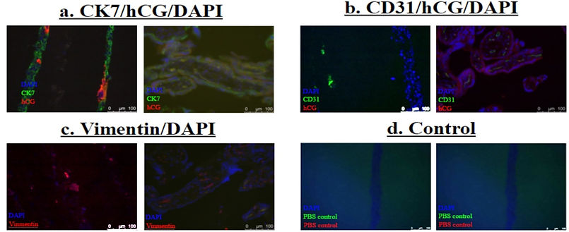
Immunofluorescence staining is conducted on our bioprinted model and 2nd trimester placenta tissue to visualise the presence of trophoblast, syncytiotrophoblast (STBs) and MSCs. By doing so we can compare the similarities between the physiological properties of our bioprinted model (shown on the left in each pair) and an actual 2nd trimester placenta tissue (shown on the right in each pair). Trophoblast, STBs, HUVEC and MSCs are present in 2nd trimester placenta. Similarly, our model also shows a presence of Trophoblast, HUVEC and MSCs. However, little to no STB was observed, this is because according to published papers JEG3 cells does not form STBs. From the staining results shown above, the model is close to a physiological placental barrier.
Anticipated Diffusion Result

By conducting literature research and using the data in published papers, we are able to come up the anticipated diffusion transfer of different substances from the maternal to fetal interface. Substances such as glucose will diffuse across the interface. We expect the concencentration of glucose in the maternal side and fetal side to be linearly related. On the other hand, substances such as insulin will not pass through the interface.